
Methods Used in Our Research
Extensive neuropathological characterization of brain tissue samples
We additionally aim to perform genetic and biochemical characterization of samples
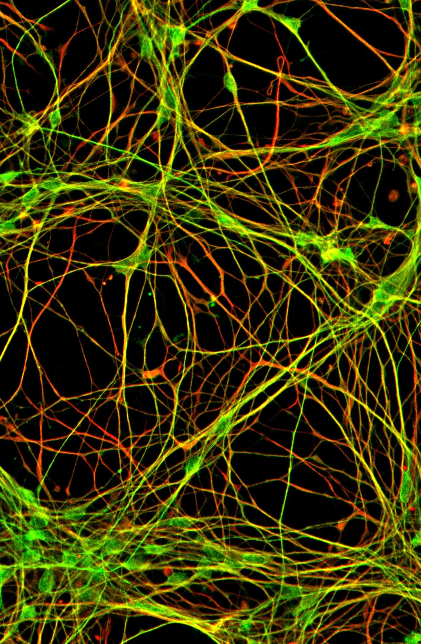
Immunohistochemistry/Immunocytochemistry: to visualize proteins in the tissue samples and evaluate using microscopy
mRNA expression methods: to evaluate RNA expression in tissue homogenates and at cellular level
3D reconstruction of scanned sections
Western blotting: to evaluate expression of proteins
Real-time quaking-induced conversion assays (RT-QuIC): to detect seeding capacity of neurodegeneration related proteins
Cell culture: to be able to perform experiments on molecular mechanisms at the cellular level
Preparation of pathological filaments from diseased brain tissue
Protein aggregation assays: to evaluate the capacity of neurodegeneration related protein to aggregate
Confocal Laser Scanning Microscopy: to evaluate co-localization of proteins
Molecular Imaging: to evaluate proteins at single-molecule based super-resolution
Electron microscopy: to evaluate the ultrastructure of protein aggregates and inclusions and cell structures